Understanding Dental X-rays (Comprehensive Guide)
Dental X-rays are a window into the hidden aspects of your oral health, revealing issues not visible during a regular exam.
They’re a safe, low-radiation tool that provides crucial insights for diagnosis and treatment.
Understanding your X-rays empowers you to participate in your dental care, ensuring you’re well informed about your oral health.
Key Highlights
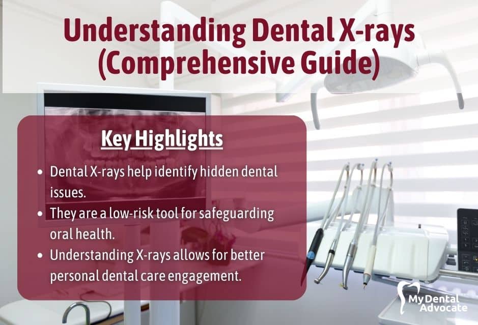
- Dental X-rays help identify hidden dental issues.
- They are a low-risk tool for safeguarding oral health.
- Understanding X-rays allows for better personal dental care engagement.
Related Posts
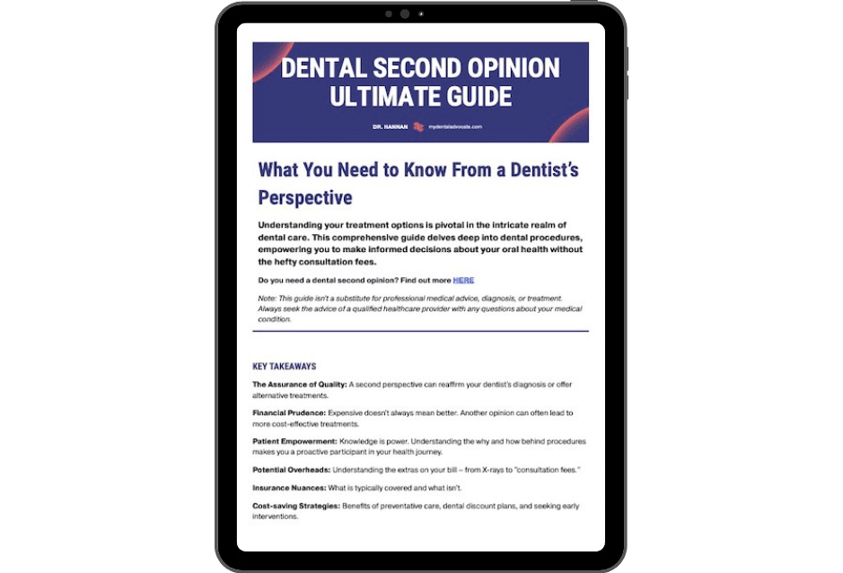
- Dental Second Opinion Ultimate Guide (Content Hub)
- Dental Crown Second Opinion (Potentially Save Thousands)
- Second Opinion Advantage (Avoiding Unnecessary Dental Work)
Understanding Dental X-rays
Dental X-rays are a crucial component in maintaining your oral health.
Need Dental Advice? Ask Dr. Hannan!
They provide invaluable insights into areas not visible during a standard dental exam, revealing the condition of your teeth, roots, jaw placement, and facial bone composition.
Dental Radiography Basics
Dental radiography refers to the process of obtaining X-ray images of your teeth and jaw.
By using low levels of radiation, these X-rays capture detailed images of your internal oral structures.
This process helps your dentist diagnose problems such as cavities or bone loss that can’t be detected with a visual examination alone.
Dental X-rays are typically quick to perform and involve minimal discomfort.
Types of Dental X-rays
There are several types of dental X-rays, each serving a different purpose:
- Bitewing X-rays show details of the upper and lower teeth in one area of your mouth. These are used to check for decay between teeth and to show how well your teeth align.
- Periapical X-rays focus on two complete teeth from root to crown. They’re used to find problems below the gum line or in the jaw, such as impacted teeth or abscesses.
- Panoramic X-rays capture an image of your entire mouth area – all teeth in both upper and lower jaws – and are useful for examining impacted teeth and planning orthodontic treatment.
How to Read a Dental X-ray
You will learn to identify various elements of your teeth and mouth structure, as well as pinpoint potential issues that may require attention.
Identifying Common Features
When you look at your dental X-ray, you’ll notice several distinct features.
Tooth enamel shows up as a bright white area due to its density, while dental fillings might appear even brighter because materials like amalgam absorb more X-rays.
Your pulp cavity, the inner part of the tooth where the nerves and blood vessels reside, is typically visible as a darker spot surrounded by the lighter enamel.
Roots of the teeth extend into the jawbone and can be seen running below the tooth crown.
They usually appear as dark channels.
Key Features
- White areas: Dense structures like enamel or fillings.
- Dark areas: Pulp cavities, spaces between teeth, and other less dense structures.
Spotting Signs of Dental Issues
Look for dark spots on the tooth, which may indicate decay or cavities.
Gum disease may be recognizable by a loss of the normally crisp border between teeth and bone, suggesting bone loss.
Additionally, changes in the shape or size of the roots or the bone surrounding the teeth might suggest an infection or another underlying condition.
Key Factors
- Dark spots on teeth: Potential cavities or decay.
- Fuzzy or irregular bone edges: Possibility of gum disease and bone loss.
- Changes in root or bone structure: May signal infection or other dental conditions.
If you spot something unusual, it’s important to consult with your dentist for a professional assessment.
To further deepen your understanding, you can explore materials on interpreting X-ray images or understanding dental X-rays.
The Importance of Dental X-rays
Dental X-rays provide a comprehensive look at your oral health, revealing issues beyond what can be seen during a regular exam.
They’re essential for accurate diagnosis and effective treatment planning.
Diagnosis & Treatment Planning
- Detect Hidden Problems: X-rays reveal unseen areas, spotting cavities, wisdom tooth issues, and potential tumors.
- Plan Treatments Effectively: X-rays help dentists tailor treatments like braces or implants, ensuring precision and timeliness.
Preventative Care Insights
- Track Developmental Changes: For children and adolescents, X-rays play a crucial role in monitoring the development of teeth and jaw structure, helping to predict and intercept potential issues early on.
- Prevent Further Decay or Damage: By identifying problems early, such as small areas of decay or signs of gum disease, X-rays support preventative care strategies that protect your oral health from progressing to more serious conditions.
Dental X-rays & Safety
When it comes to dental X-rays, your safety is paramount.
This section will cover the aspects of radiation you’re exposed to during an X-ray and the safety measures in place to protect you.
Radiation Exposure
Your exposure to radiation during a dental X-ray is extremely low.
Dental X-rays emit a type of radiation that helps capture detailed images of your teeth and bones, which are crucial for diagnosis and treatment planning.
The level of radiation is measured in millirems (mrem), and a typical dental X-ray gives off approximately 0.5 mrem per image.
Safety Measures & Precautions
Dentists take a range of safety measures to further minimize your exposure to radiation:
- Lead Aprons: This protective shield helps safeguard your body, especially your vital organs, from unnecessary exposure.
- Thyroid Collars: A specialized collar that protects your thyroid gland from X-ray radiation.
Additionally, precautions include using the latest technology, such as digital X-rays, which can reduce radiation exposure significantly compared to traditional film X-rays.
Your dentist will also adhere to the principle of ALARA (As Low As Reasonably Achievable), ensuring that X-rays are used only when necessary to minimize your cumulative radiation dose.
For concrete data on safety standards and exposure, refer to The Super Dentists’ article on Dental X-Rays and Safety, and for a deeper look into types of dental X-rays and their specific uses, see Cleveland Clinic’s resource on Dental X-Rays: Types, Uses & Safety.
Preparing for Dental X-rays
Getting a dental X-ray is a routine process that helps your dentist assess your oral health.
Before the Appointment
- Research: Familiarize yourself with the types of dental X-rays to understand what you might need.
- History: Compile a list of your current medications and past dental records if you visit a new dentist.
- Questions: Have any concerns or questions written down to ask your dentist before the procedure begins.
During the Procedure
- Jewelry: Remove any earrings, necklaces, or facial piercings to avoid interference with the X-ray images.
- Following Instructions: Stay still, as movement can blur the images and might necessitate retakes.
Remember, these steps are designed to give you peace of mind and ensure clear, accurate results from your X-ray.
Interpreting X-ray Results
When your dentist shows you dental X-rays, they serve as a vital tool for identifying hidden dental conditions that require attention.
Let’s break down how these images are analyzed and communicated to you.
Professional Analysis
After your dental X-rays are taken, the dentist or a dental radiologist carefully examines them for a range of issues.
It is essential to recognize that different types of X-rays reveal various aspects of your oral health.
Bitewing X-rays are ideal for detecting cavities between teeth, while periapical X-rays show the entire tooth from the crown to the root, offering a clear view of the supporting bone structure.
The dentist looks for changes in bone density, the condition of your tooth roots, and signs of infection or decay that are not visible during a standard examination.
Patient Communication
Once the X-ray analysis is complete, your dentist will discuss the findings with you.
Expect a straightforward conversation where they will point out any areas of concern, such as cavity formations, indications of periodontal disease, or impacted teeth.
Your understanding is critical, so they might use terms like “dark spots” for cavities or “fuzzy areas” for potential bone loss.
Your dentist’s goal is not only to inform you of the present condition but also to involve you in deciding on the appropriate treatment.
Advances in Dental Imaging
In the realm of dental care, imaging technology has made leaps and bounds, enhancing both diagnostic capabilities and patient comfort.
Understanding modern dental imaging technology can help you make informed decisions about your oral health.
Digital X-ray Technology
Digital X-rays have revolutionized dental diagnostics, offering benefits like reduced radiation exposure and immediate image processing.
Unlike traditional film X-rays, you can view digital X-rays on a computer screen right after they’re taken.
This technology also makes it easier for your dentist to store and share your X-ray images, should you need to consult with a specialist or transfer records.
Benefits of Digital X-ray Technology
- Reduced radiation exposure compared to traditional film X-rays
- Fast image processing for immediate results
- Enhanced image quality for better diagnosis
- Easy sharing and storage
3D Imaging & CBCT
Cone Beam Computed Tomography (CBCT) is a form of 3D imaging that provides your dentist with a comprehensive view of your teeth, soft tissues, nerve pathways, and bone in a single scan.
This detailed perspective is particularly useful for complex procedures such as dental implant planning, orthodontic treatments, and root canal therapy.
Uses of 3D Imaging and CBCT
- Accurate planning for dental implants
- Detailed analysis for orthodontics
- Precise mapping for root canal treatments
- Improved detection of dental issues
With 3D imaging and CBCT, your dental visits can become more efficient, and your treatment outcomes can improve due to the high level of detail these scans provide.
MDA Verification Report
What is the My Dental Verification Report, and why is it important?
Our board-certified dentists will review your case and provide an unbiased second opinion on how you should move forward with your proposed dental treatment.
We respect all dentists suggested treatment; however, we desire clarity for your dental needs.
You should understand what dental work is necessary to move forward with treatment confidently.

Highlights
- Avoid unnecessary dental treatment
- Potentially save you thousands of dollars
- Information reviewed privately and securely
- Unbiased assessment that brings you peace of mind
- Quick response time to review the report at your convenience
- Clarity and confidence moving forward with dental treatment
- Completed from the comfort of your own home
What’s Involved?
With My Dental Advocate, patients of all ages now have an unbiased, expert second opinion to securely review their proposed dental treatment and gain the knowledge to improve their oral health.
Step 1: Complete Questionnaire
The questionnaire includes a brief medical history, oral hygiene habits, and an opportunity to explain your dental concerns.
Step 2: Upload X-rays
Contact your provider to request your x-rays or take a photo of them at the office. You can also upload dental photos, current treatment plans, periodontal probings, and other supporting documents.
Step 3: Submit Payment
We will privately send your information to a board-certified dentist who will securely assess your case and provide a detailed second opinion for your proposed dental work.
Step 4: Receive MDA Verification Report
We will email your MDA Verification Report second opinion within hours for review at your convenience and from the comfort of your home.
Frequently Asked Questions (FAQ)
My Experience & Expertise
As a dentist, I can’t stress enough the importance of dental X-rays.
They are not just images, but a crucial part of your dental health journey.
They allow us to see beyond the surface, catch problems early, and tailor treatments effectively.
Embracing this technology means taking a proactive step towards maintaining a healthy, happy smile.
Need a second opinion? We can help! Learn more. Knowledge is power when cultivating healthy dental habits. The more informed you are, the better positioned you’ll be to prevent avoidable and potentially costly dental procedures for you and your family. Watch for future blog posts, where we’ll continue sharing important information, product reviews and practical advice!

About the Author
Dr. Matthew Hannan, also known as “Dr. Advocate,” is a board-certified dentist on a mission to provide accurate dental patient education. He attended Baylor University before completing dental school at UT Health San Antonio School of Dentistry. He now lives in Arizona with his beautiful wife and 4 kids. Dr. Hannan believes everyone should access easy-to-read dental resources with relevant, up-to-date dental research and insight to improve their oral health.

Connect with Dr. Hannan!

Does Filtering Water Remove Fluoride? (2024 Update)
Fluoride is a mineral added to drinking water for decades to improve dental health. However, only some enjoy fluoride in their drinking water, and many wonder if filtering their water can remove this mineral…
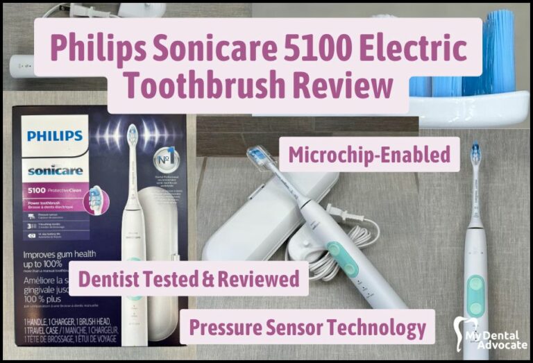
Sonicare 5100 Electric Toothbrush (Full Review)
Looking for a sparkling smile and improved oral health? Search no more! Introducing the Philips Sonicare ProtectiveClean 5100, an electric toothbrush that’s both powerful and customizable, designed to provide an unparalleled deep…
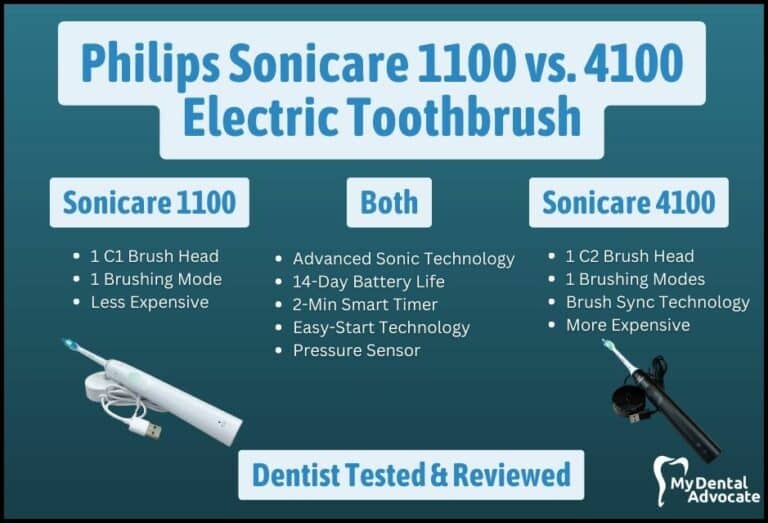
Philips Sonicare 1100 vs. 4100 Electric Toothbrush Review 2024
Brushing your teeth has never been more advanced! Dive into our detailed comparison of Philips Sonicare 1100 vs. 4100 electric toothbrushes. Uncover the nuances and discover which brush offers you the ultimate oral care experience. Let the battle of the bristles begin!
Gain Clarity with Our FREE Second Opinion Guide
Receive clear, expert second opinions online within 48 hours. Start today!
Product Reviews
Our 250+ dental product reviews (and counting), curated by an experienced dentist, are the most comprehensive online.
Toothbrush Genie
State-of-the-art chatbot designed to help you discover your perfect toothbrush in just a few simple steps!
Cavity Risk Assessment
Cutting-edge digital tool designed to evaluate your individual cavity risk based on your responses to a series of questions.
Gum Disease Assessment
Discover your gum disease risk with our quick and engaging 6-question assessment!
